
-
摘要: 为研究氧化铈刻蚀对于金刚石表面形貌的影响,将金刚石单晶与氧化铈粉末以质量比1:5的比例混合,并在N2气氛下对金刚石进行刻蚀处理。通过对刻蚀后金刚石的表面形貌、表面刻蚀深度、物相组成的表征与分析探究氧化铈粉末刻蚀对于金刚石表面形貌的影响。利用铜基结合剂金刚石试样的抗弯强度评估刻蚀对于金刚石与结合剂之间把持力的影响。结果表明:氧化铈能成功对金刚石单晶表面进行选择性刻蚀。随着温度的升高,金刚石各个晶面的刻蚀深度加深;在相同条件下,氧化铈对金刚石(100)面的刻蚀程度大于金刚石(111)面。当刻蚀温度为900 ℃时,金刚石(111)面刻蚀深度为753.23 nm,(100)面刻蚀深度为1.60 μm。在N2气氛下,900 ℃氧化铈刻蚀后的金刚石单颗粒抗压强度低于未加氧化铈刻蚀剂刻蚀的金刚石抗压强度,但是高于在空气气氛下未加氧化铈刻蚀剂刻蚀的金刚石以及在空气中直接氧化铈刻蚀的金刚石抗压强度。与未处理金刚石相比,氧化铈刻蚀后的铜基结合剂金刚石试样的抗弯强度有较大提升。Abstract: To investigate the effect of cerium oxide (CeO2) etching on the surface morphology of diamond, the diamond single crystals and cerium oxide powders were mixed in the ratio of 1:5 by mass and etched under the N2 atmosphere. The effect of cerium oxide powder etching on the surface morphology of diamond was investigated by characterizing and analyzing the surface morphology, etching depth, and phase composition of the diamond after etching. The effect of etching on the holding force between the diamond and bond was also evaluated by the flexural strength of the diamond specimens with the copper-based bond. The results show that the cerium oxide can successfully etch the surface of diamond single crystal selectively. As the temperature increases, the etching depth of each crystalline surface of diamond deepens, and under the same conditions, the etching depth of cerium oxide on the diamond (100) surface is greater than that on the diamond (111) surface. At the etching temperature of 900 ℃, the etching depth of diamond (111) surface is 753.23 nm and that of (100) surface is 1.60 μm. The compressive strength of the diamond single crystal etched by cerium oxide at 900 ℃ in the N2 atmosphere is lower than that of the diamond etched without etchant in the N2 atmosphere, but higher than that of the diamond etched without etchant in the air atmosphere and the diamond etched by cerium oxide directly in the air. Compared with the untreated diamonds, the flexural strength of the copper-bonded diamond specimens after cerium oxide etching is improved.
-
Key words:
- diamond single crystal /
- cerium oxide /
- surface morphology /
- anisotropic etching
-

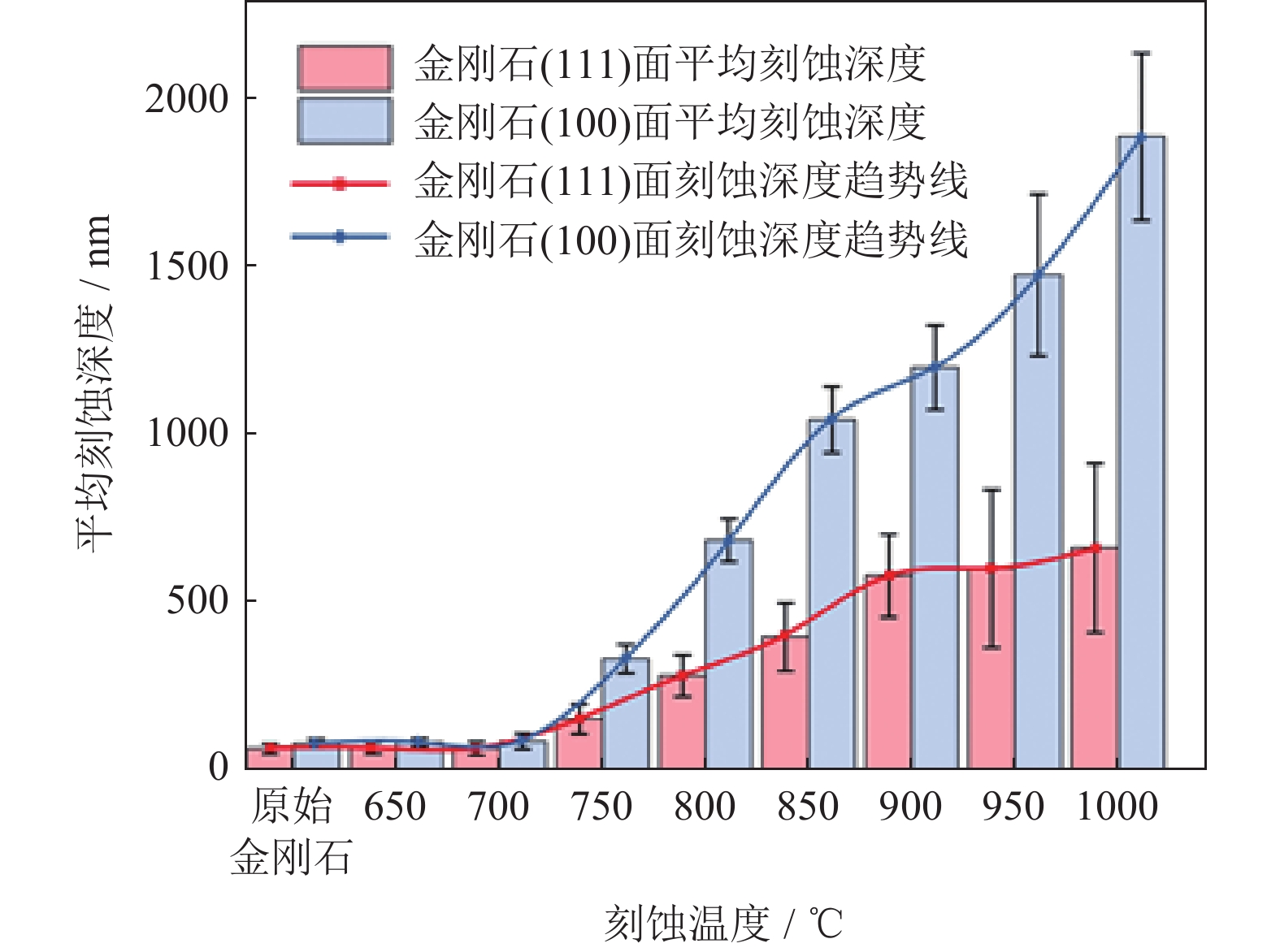
图 1 不同刻蚀温度金刚石表面刻蚀深度
Figure 1. Etching depth of the diamond surface at the different etching temperatures

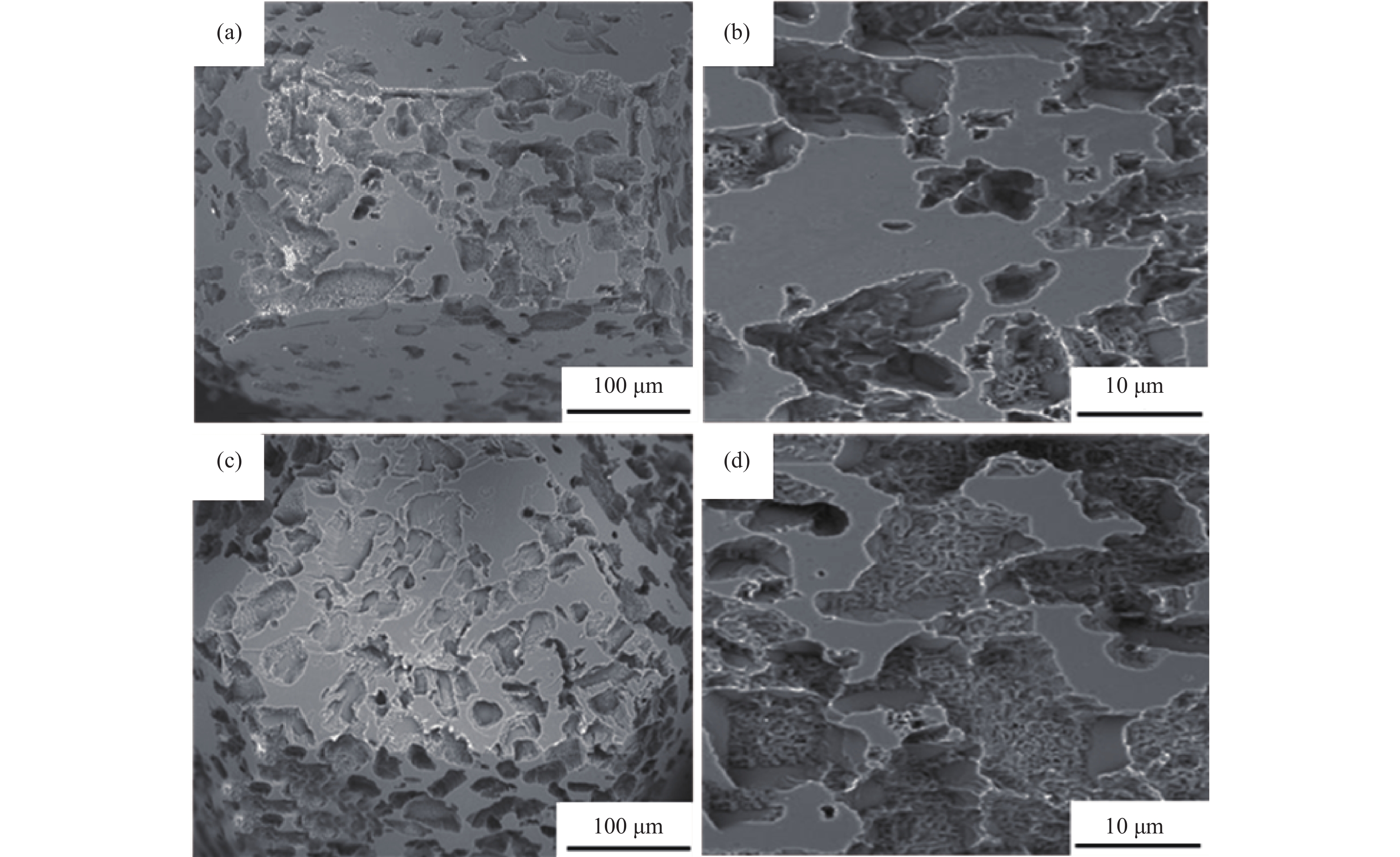
图 2 950 ℃和1000 ℃刻蚀金刚石表面形貌:(a)、(b)950 ℃;(c)、(d)1000 ℃
Figure 2. Etching morphology of the diamond surface at 950 ℃ and 1000 ℃: (a), (b) 950 ℃; (c), (d) 1000 ℃

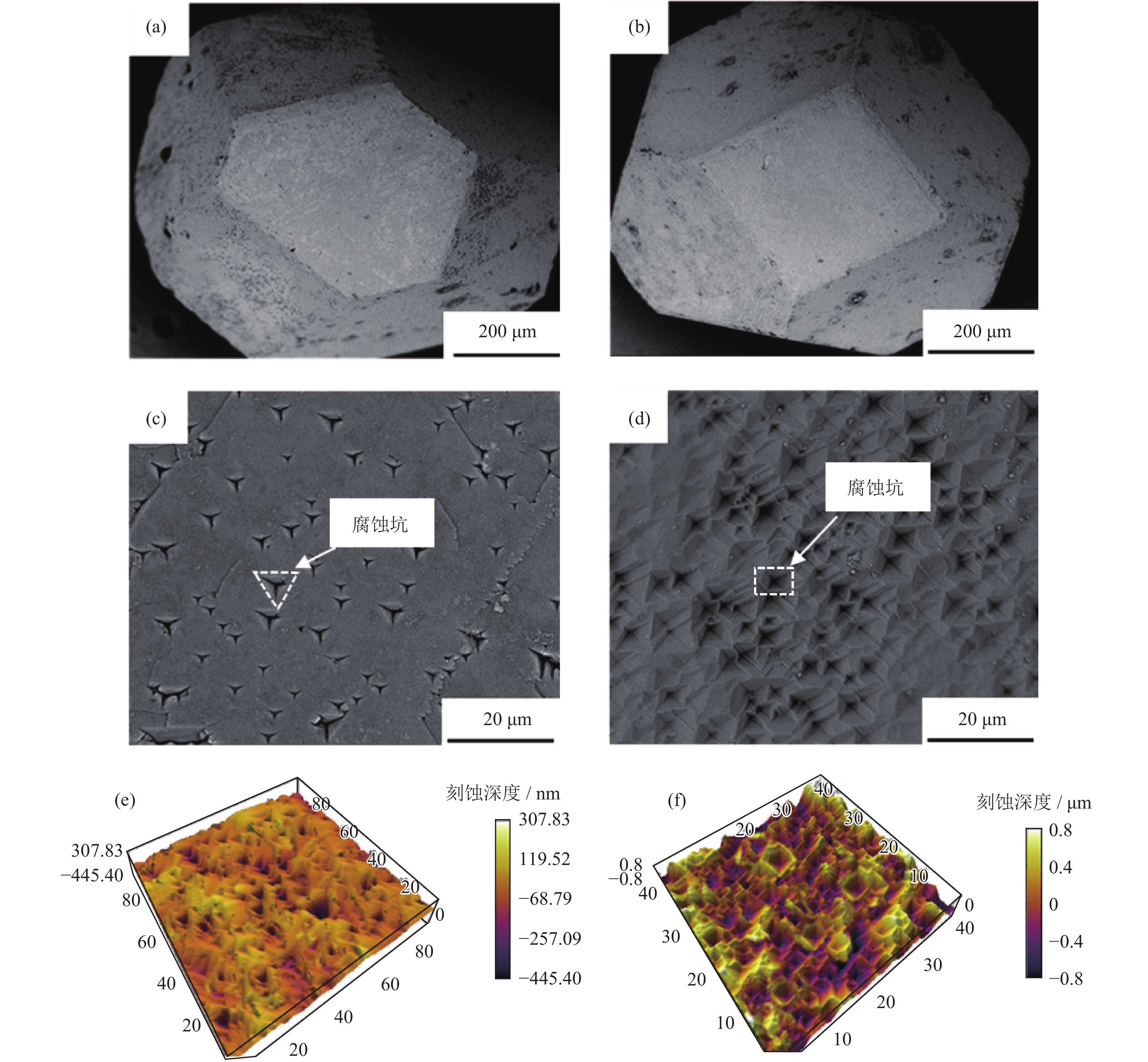
图 3 900 ℃刻蚀后金刚石表面形貌:(a)、(c)、(e)(111)面;(b)、(d)、(f)(100)面
Figure 3. Surface morphology of the diamond after etching at 900 ℃: (a), (c), (e) (111) surface; (b), (d), (f) (100) surface

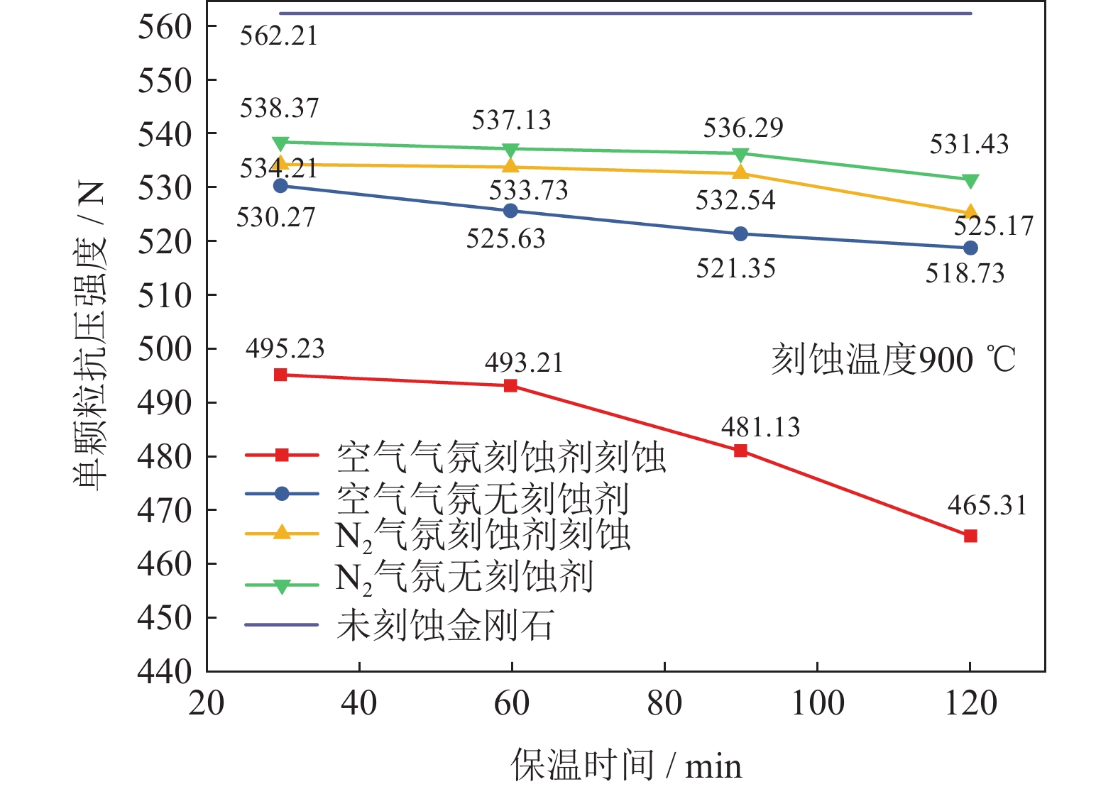
图 4 不同刻蚀条件下金刚石单颗粒抗压强度
Figure 4. Compressive strength of the diamond single crystal in the different etching condition

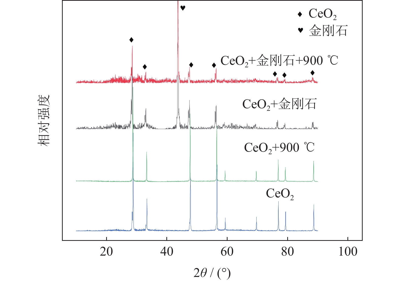
图 5 原始氧化铈粉末、氧化铈与金刚石混合物、900 ℃氧化铈粉末、900 ℃氧化铈与金刚石混合物的X射线衍射图谱
Figure 5. XRD spectra of the original CeO2 powders, CeO2 and diamond mixture, CeO2 powders at 900 ℃, and CeO2 and diamond mixture at 900 ℃

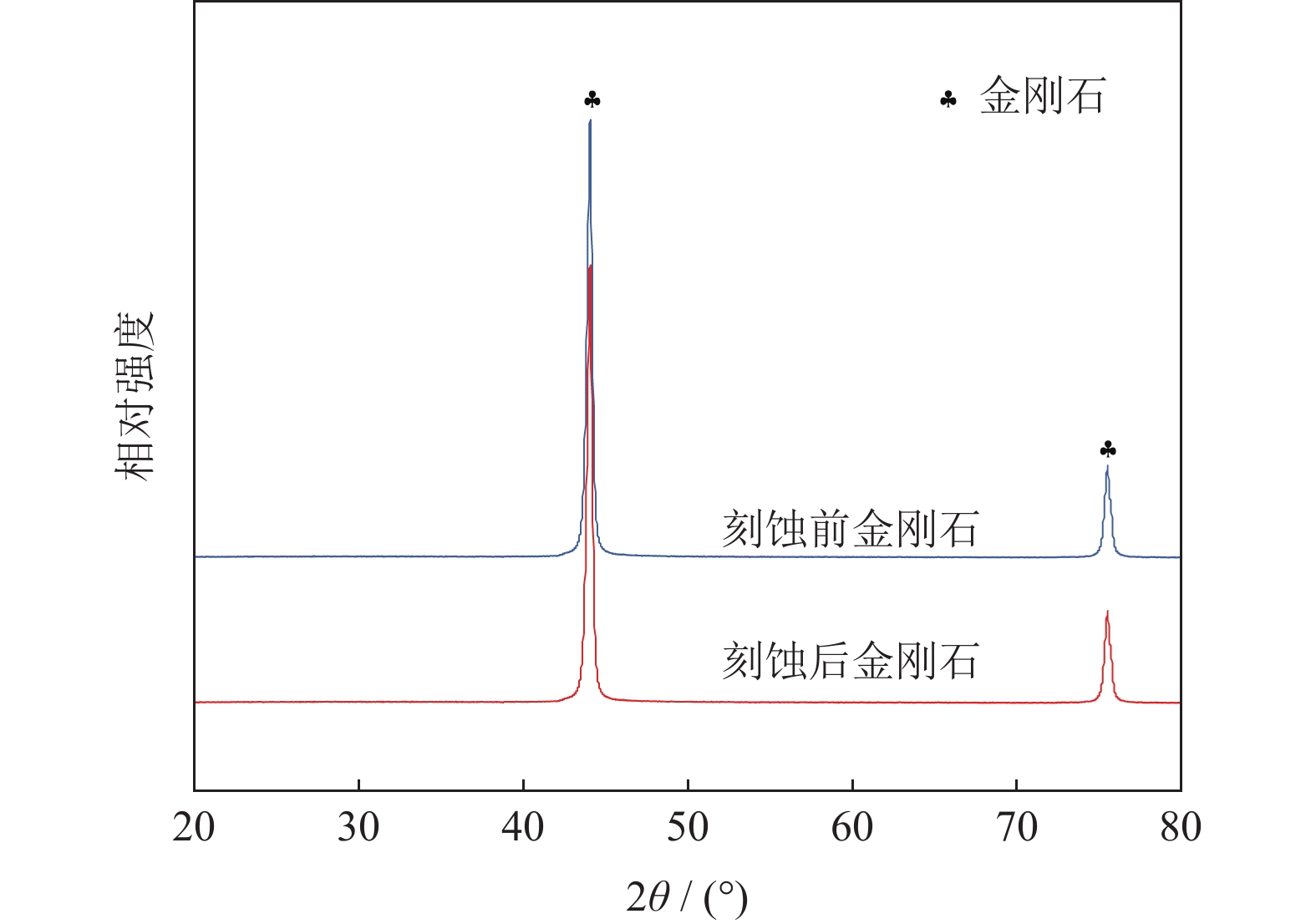
图 6 900 ℃刻蚀前后金刚石单晶X射线衍射图谱
Figure 6. XRD spectra of the diamond single crystal before and after etching at 900 ℃

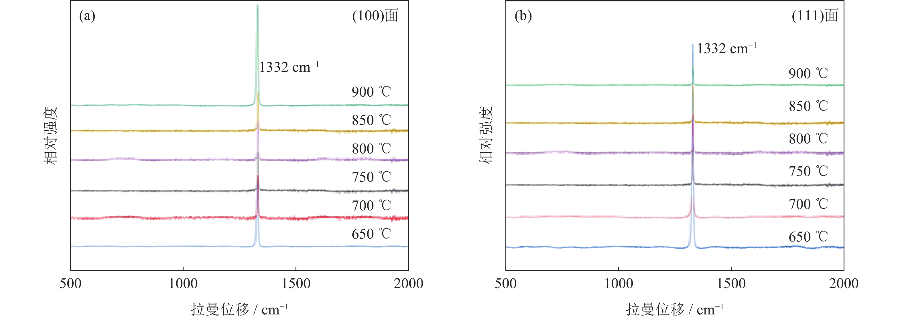
图 7 金刚石刻蚀处拉曼图谱分析:(a)(100)面;(b)(111)面
Figure 7. Raman spectrum analysis of the diamond etching: (a) (100) surface; (b) (111) surface

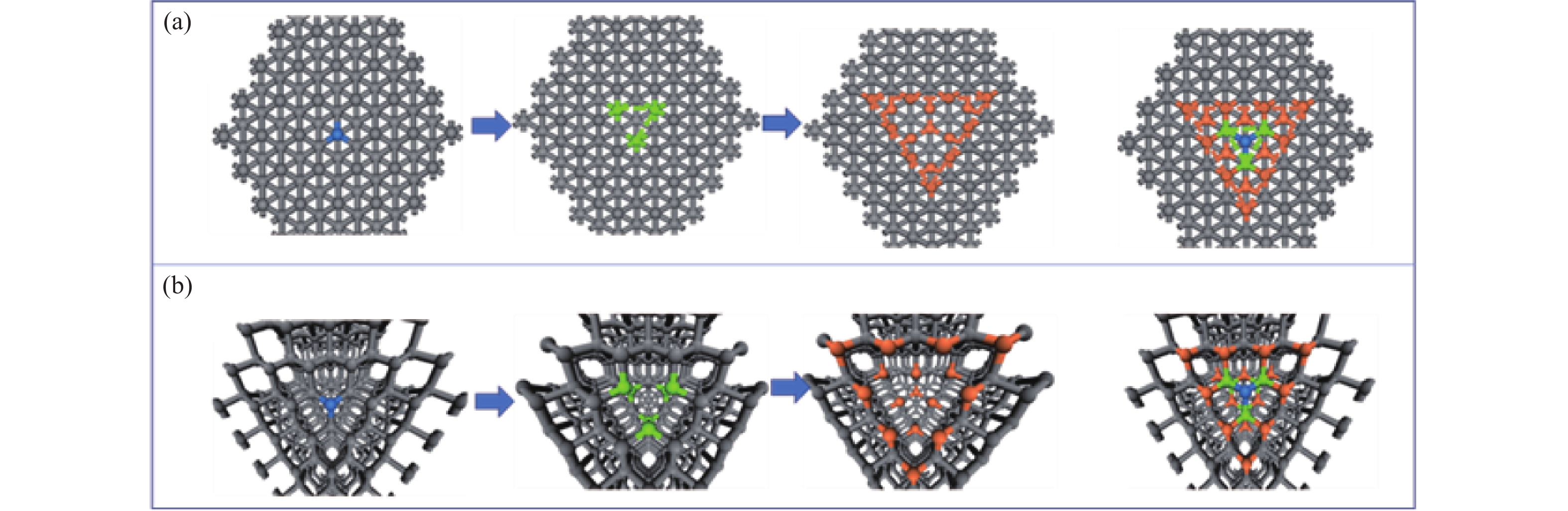
图 8 金刚石(111)面刻蚀原理示意图:(a)脱落原子排布平面示意图;(b)脱落原子排布立体示意图
Figure 8. Schematic diagram of the etching principle in diamond (111) surface: (a) schematic diagram of the dropped atom arrangement ; (b) three-dimensional schematic diagram of the fallen atom arrangement

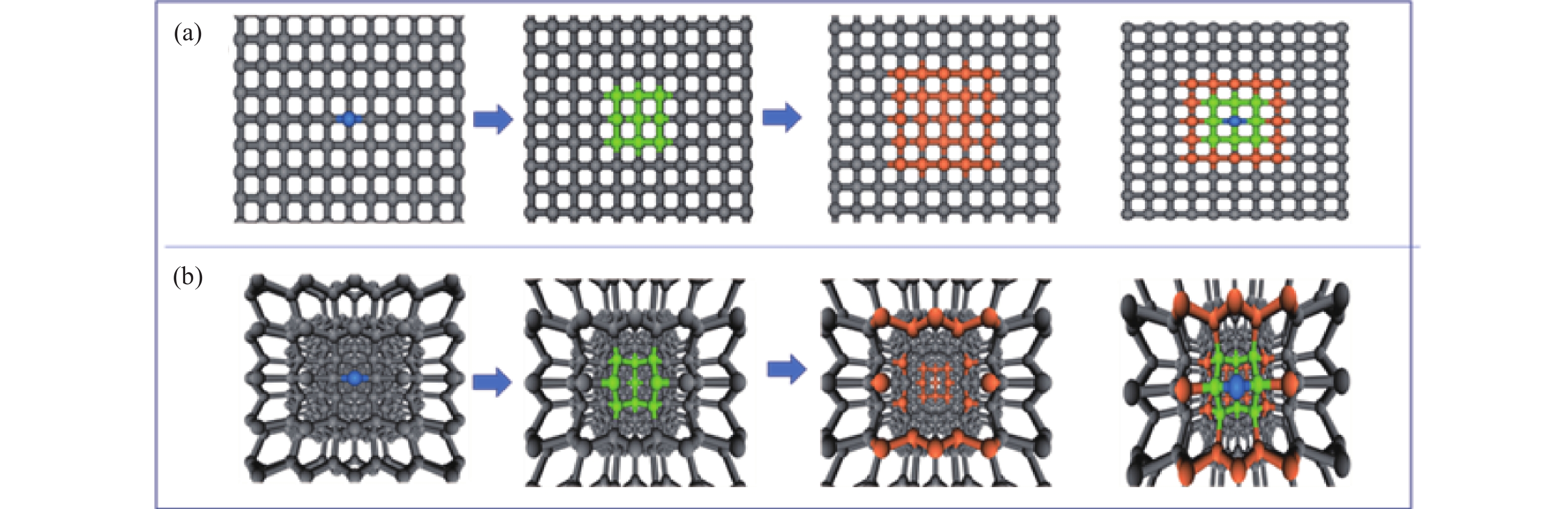
图 9 金刚石(100)面刻蚀原理示意图:(a)脱落原子排布平面示意图;(b)脱落原子排布立体示意图
Figure 9. Schematic diagram of the etching principle in diamond (100) surface: (a) schematic diagram of the dropped atom arrangement ; (b) three-dimensional schematic diagram of the fallen atom arrangement
-
[1] Zhu X Y, Kwok S Y, Yuen M F, et al. Dense diamond nanoneedle arrays for enhanced intracellular delivery of drug molecules to cell lines. J Mater Sci, 2015, 50(23): 7800 doi: 10.1007/s10853-015-9351-z [2] Scorsone E, Gattout N, Rousseau L, et al. Porous diamond pouch cell supercapacitors. Diamond Relat Mater, 2017, 76: 31 doi: 10.1016/j.diamond.2017.04.004 [3] Khanaliloo B, Mitchell M, Hryciw A C, et al. High-Q/V monolithic diamond microdisks fabricated with quasi-isotropic etching. Nano Lett, 2015, 15(8): 5131 doi: 10.1021/acs.nanolett.5b01346 [4] Li Y Y, Wan L, Wang J S, et al. Influence of temperature on etching and foaming behavior of synthetic diamond powder by iron-based pre-alloyed powders. Mater Rev, 2017, 31(14): 113 doi: 10.11896/j.issn.1005-023X.2017.014.024李颖颖, 万隆, 王俊沙, 等. 温度对铁基预合金粉腐蚀泡沫化人造金刚石微粉的影响. 材料导报, 2017, 31(14): 113 doi: 10.11896/j.issn.1005-023X.2017.014.024 [5] Morofushi Y, Matsushita H, Miki N. Microscale patterning of single crystal diamond by thermochemical reaction between sidero-metal and diamond. Precis Eng, 2011, 35(3): 490 doi: 10.1016/j.precisioneng.2011.03.003 [6] Masatsugu N, Kazuhiro N, Hiraku T, et al. Anisotropic diamond etching through thermochemical reaction between Ni and diamond in high-temperature water vapour. Sci Rep, 2018, 8(1): 6687 doi: 10.1038/s41598-018-25193-2 [7] Enriquez J I, Muttaqien F, Michiuchi M, et al. Oxidative etching mechanism of the diamond (100) surface. Carbon, 2021, 174: 36 doi: 10.1016/j.carbon.2020.11.057 [8] Li L Y, Chen X, Zhang W, et al. Characterization and formation mechanism of pits on diamond {100} face etched by molten potassium nitrite. Int J Refract Met Hard Mater, 2018, 71: 129 doi: 10.1016/j.ijrmhm.2017.11.011 [9] Xiao C J, Dou Z Q, Zhu Z D. Characterization and formation mechanism of surface morphology on diamond etched by Fe2O3 powder. Mater Rep, 2020, 34(14): 14045 doi: 10.11896/cldb.19070180肖长江, 窦志强, 朱振东. 氧化铁刻蚀金刚石表面形貌的表征及形成机理. 材料导报, 2020, 34(14): 14045 doi: 10.11896/cldb.19070180 [10] Wang J S, Wan L, Chen J, et al. Micropatterning of diamond crystallites via cobalt-catalyzed thermochemical etching. J Mater Sci, 2017, 52(2): 709 doi: 10.1007/s10853-016-0365-y [11] Wang J S, Wan L, Chen J, et al. Anisotropy of synthetic diamond in catalytic etching using iron powder. Appl Surf Sci, 2015, 346: 388 doi: 10.1016/j.apsusc.2015.04.022 [12] Wang J S, Wan L, Chen J, et al. Surface patterning of synthetic diamond crystallites using nickel powder. Diamond Relat Mater, 2016, 66: 206 doi: 10.1016/j.diamond.2016.04.010 [13] Wei Y J, Anand L. Grain-boundary sliding and separation in polycrystalline metals: application to nanocrystallinefcc metals. J Mech Phys Solids, 2004, 52(11): 2587 doi: 10.1016/j.jmps.2004.04.006 [14] Pehrsson P, Mercer T W, Chaney J A. Thermal oxidation of the hydrogenated diamond (100) surface. Surf Sci, 2002, 497(1-3): 13 doi: 10.1016/S0039-6028(01)01677-6 [15] Dou Z Q, Xiao C J, Ren F H, et al. Effect of holding time on etched diamond single crystal by manganese oxide. Powder Metall Technol, 2019, 37(5): 344窦志强, 肖长江, 任付豪, 等. 保温时间对锰氧化物刻蚀金刚石单晶的影响. 粉末冶金技术, 2019, 37(5): 344 [16] Xiao C J, Chen Y G, Li X L, et al. Method and mechanism analysis of improving the holding force between Ni-coated diamond and Cu-matrix bonding. Powder Metall Technol, 2020, 38(1): 25肖长江, 陈贻光, 栗晓龙, 等. 镀 Ni 金刚石与铜基结合剂间把持力的提高方法和机理分析. 粉末冶金技术, 2020, 38(1): 25 -

 下载:
下载:

 点击查看大图
计量
- 文章访问数: 569
- HTML全文浏览量: 276
- PDF下载量: 55
- 被引次数: 0
